


Knee mensicus/ cartilage treatment
Keyhole knee surgery (knee arthroscopy)
Knee ligament repair (ACL repair)
PRP knee injections
Hyaluronic acid knee injections
Robotic assisted MAKO/NAVIO Knee Replacement
Robotic knee surgery explained
Watch Richard's surgery and recovery after robotic knee replacement
Watch Tibor recover from a computer guided partial knee replacement
Knee cartilage/Menicus repair
Arthroscopic Meniscus Repair (AMR) is a keyhole surgical procedure through small 1cm incisions. Its aim is to achieve healing of meniscal tissue which has torn and is carried out to reduce pain and slow degeneration (wear and tear) of the knee joint.
To understand why AMR needs to be performed, it is first necessary to understand the anatomy and damage that takes place.
The menisci are two C shaped pieces of fibrocartilage that line your knee joint, providing cushioning and stability between the femur (thigh bone) and tibia (shin bone). They increase the contact area between the two bones and hence reduce abnormal stress forces during knee motion.
The medial meniscus is more commonly injured and is located on the inner aspect of your knee, while the lateral meniscus is located at the outer aspect of the knee.
Either or both menisci can be torn during activities that put excess pressure, twisting and bending forces on the knee joint such as football, netball, tennis and skiing.
Older patients are more likely to tear their meniscus with less strenuous activity as meniscal tissue degenerates with age making it weaker and less pliable.
The benefits of undergoing AMR include reduced pain levels, cessation of any unwanted mechanical symptoms from the knee such as locking or giving way and slowing down future osteoarthritis and degeneration to the joint.
The risk of complication is present with every surgical procedure but is very rare with AMR at around 0.1 – 1%, the most common being infection and deep vein thrombosis and/or pulmonary embolism.
The procedure is normally performed under general anaesthetic, and usually takes between 20 minutes to 1 hour. When you wake up, you should not be in too much discomfort as local anaesthetic is injected into the knee before the procedure ends, and effective painkillers are prescribed for you to take at home for the first 2-5 days if required.
Patients are allowed to walk the same day and return home, normally with crutches for comfort, bandaging over the knee and sometimes a special knee support (knee brace) to wear for a few weeks.
At home you should keep your leg elevated when resting, with an ice pack placed on the knee for one or two days to help reduce any swelling and continue with the gentle exercise protocol your physiotherapist and surgeon have advised.
This is important to prevent the knee from getting stiff and also helps build up your thigh (quadriceps, hamstrings) and calf muscles (gastrocnemius, soleus) for a speedy recovery.
What is Arthroscopic Meniscus Repair (AMR)?
Arthroscopic Meniscus Repair (AMR) involves repairing the torn meniscus using an arthroscope and other surgical tools. An arthroscope is a thin, fibreoptic high definition camera with a similar diameter to a drinking straw. The miniature camera is attached to a monitor similar to a TV screen that allows the surgeon to see the inside of the knee without needing to make a large incision.
Because the arthroscope is inserted into the knee joint through a small 1cm incision (portal), it has certain advantages when compared to open surgery through larger incisions including:
less pain after the operation, lower risk of infection, reduced hospitalisation time, quicker recovery and return to function.
The procedure can be split into two types:
1) meniscus repair,
2) partial meniscectomy (removal of some of the damaged meniscus)
Why is AMR performed?
Surgery is not always necessary for a meniscus tear. Non operative treatment in the form of physiotherapy should always be pursued as a first line option.
The decision to treat the meniscus tear surgically depends on the frequency of your unwanted symptoms, which include focal pain and tenderness over the knee, locking (where you are unable to straighten your knee), a feeling of instability (knee giving way), and difficulty kneeling, squatting or twisting and turning.
The decision to repair the meniscus versus remove the damaged part depends on the type of meniscus tear and the location of the tear within the meniscus.
Only the peripheral part of the meniscus has a good blood supply, and as blood is required for healing, generally these peripheral tears respond best to repair with stitches.
Patient factors such as younger age ( <40 years old), no arthritis in the joint, those of a healthy weight (BMI<30) and those willing to engage in pre and post-operative physiotherapy also have a higher rate of success with AMR.
Importantly, the overarching principle of arthroscopic meniscus surgery is that as much functioning meniscus should be kept as possible, this is why at all time , where possible it is in the best interests of the patient to repair viable torn meniscus.
If performed successfully, AMR will preserve as much of the meniscus as possible, which slows joint degeneration, increase knee stability during complex activities and exercise and can significantly reduce pain.
The alternative to AMR is known as Arthroscopic Partial Meniscectomy (APM)and is the more frequently performed surgery in the treatment of Meniscal tears.
It means removal of the torn piece of meniscus and is performed in all cases where the meniscus cannot be repaired for technical reasons or because the torn piece has no viable blood supply and repairing it would lead to inevitable failure and re-tear in the weeks following surgery.
APM is also more widely performed by orthopaedic surgeons as it was traditionally the only method of surgically treating meniscal tears and is technically easier to perform.
It is important you are treated by a surgeon who is comfortable and experienced in all the available treatment options so the correct one is chosen for you.
Anaesthesia
AMR can be performed either under general anaesthesia (asleep attached to a ventilator with a tube in your windpipe to help you breathe) or spinal anaesthesia, where local anaesthetic is injected close to the spine to numb you from the waist down for about 3 hours.
Additionally, local anaesthetic is injected into the incision sites and the knee joint cavity at the end of the procedure to help ease any post-operative discomfort.
Current literature shows that spinal anaesthesia has some evidence for improved mortality and morbidity (less complications) when compared with general anaesthesia, in particular for longer orthopaedic operations such as hip and knee replacement surgery, or surgery to fix broken hip bones.
Pain Control
During the surgery, local anaesthetic such as bupivacaine will minimise the pain you experience. As AMR involves soft tissue surgery, it is moderately painful. To reduce pain post-operatively, oral low dose opioids such as codeine phosphate and non-steroidal anti-inflammatory drugs (NSAIDs) such as ibuprofen are sometimes prescribed for a few days and can be taken as required. It is important to try and avoid opioids if possible and certainly not take them regularly for more than 1 week as they are addictive and have unwanted side effects such as constipation and nausea.
Will I need to do any preparation?
Pre assessment clinic and checks
You will be booked in to attend a pre-assessment clinic prior to your surgery. The aim of the appointment is to assess your general health and assess how safe it is for you to have the surgery and anaesthetic. You will be asked about any medication and supplements you may be taking, as some of these may increase your chance of bleeding or affect the anaesthesia. Warfarin and Rivaroxaban are examples of blood thinners which your doctor will advise you to stop taking prior to the surgery.
A bespoke after care plan will also be made to ensure that you are well prepared to get home safely and be able to cope following the surgery.
The surgical team and your consultant will explain the risks and benefits associated with AMR surgery, after which point you will be asked to sign a consent form for the surgery to go ahead. You should be given adequate time to rethink your decision before the date of the surgery should you wish to ask any more questions or change your mind.
The day of surgery
On the day of surgery, you will need to have fasted for 6 hours minimum (no food or drink).
You will be met by your surgeon and anaesthetist who will go over any other questions you might have, and they will mark the site of the surgery with a marker pen. You will get into a hospital gown and be admitted by one of the nurses or health care assistants.
You will be escorted to the operating theatres on foot or a trolley, initially to the anaesthetic room. Once you enter the anaesthetic room, the anaesthetic doctor and a healthcare professional called an operating department practitioner (ODP) who assists the anaesthetist will begin the process of general anaesthetic. This begins with the placement of a small plastic tube into the vein in the back of your hand so they can inject the anaesthetic medicine which puts you into a deep sleep. They will then place a larger breathing tube (ET tube) into your windpipe just past your vocal cords carefully and attach it to a ventilator which helps regulate and monitor your breathing during the surgery.
Inside the operating theatre
You will then be wheeled into the operating theatre to be positioned face up on the operating table, lying on your back. A tourniquet cuff may or may not be attached to your thigh to limit blood flow to the knee, depending on the surgeon’s preference. Further, the knee to be operated on will be thoroughly cleaned with antiseptic to prevent infection and sterile surgical drapes will then be placed onto that knee, exposing the cleaned surgical site, by the surgeon and theatre nurse who are wearing sterile gloves and theatre gowns.
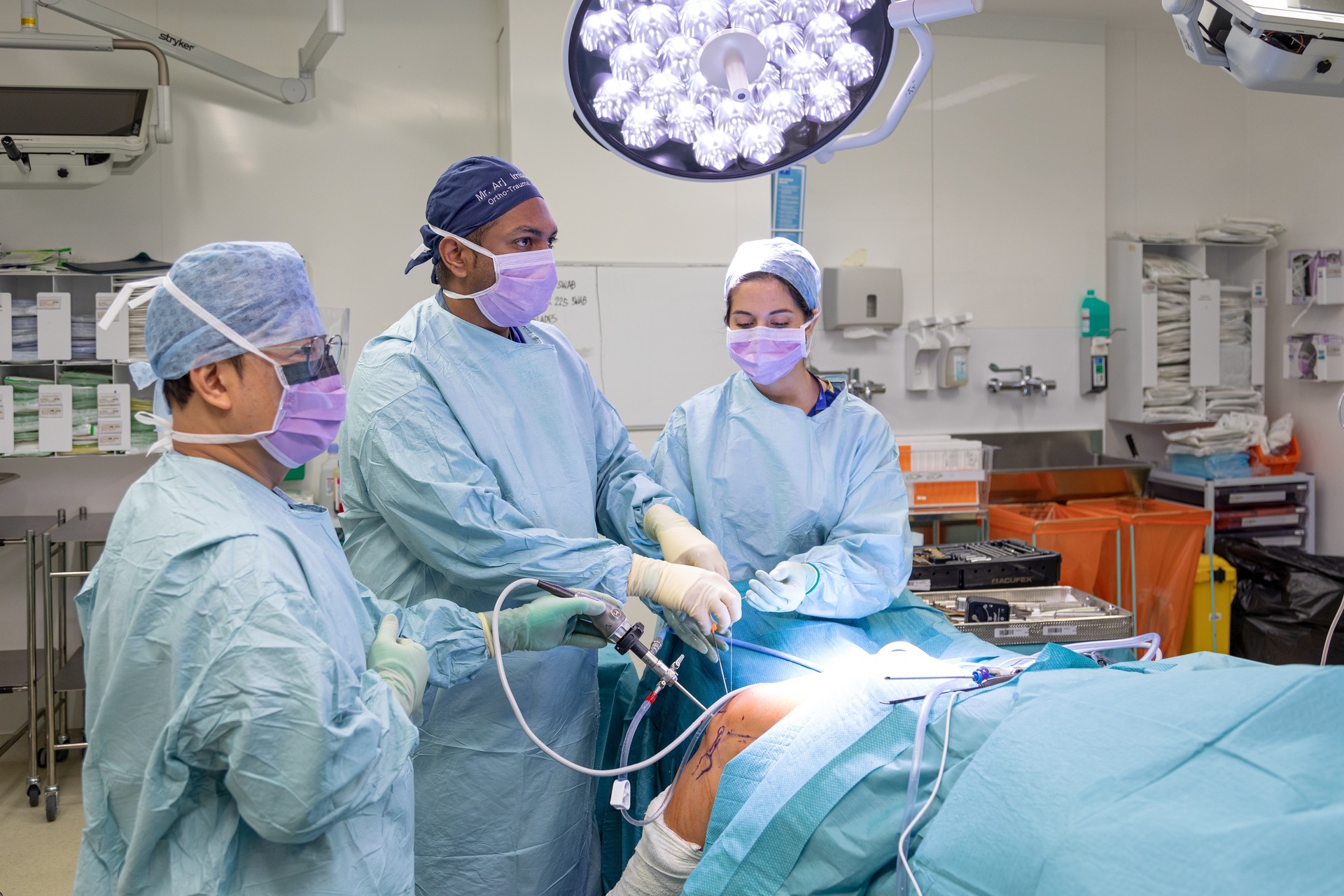
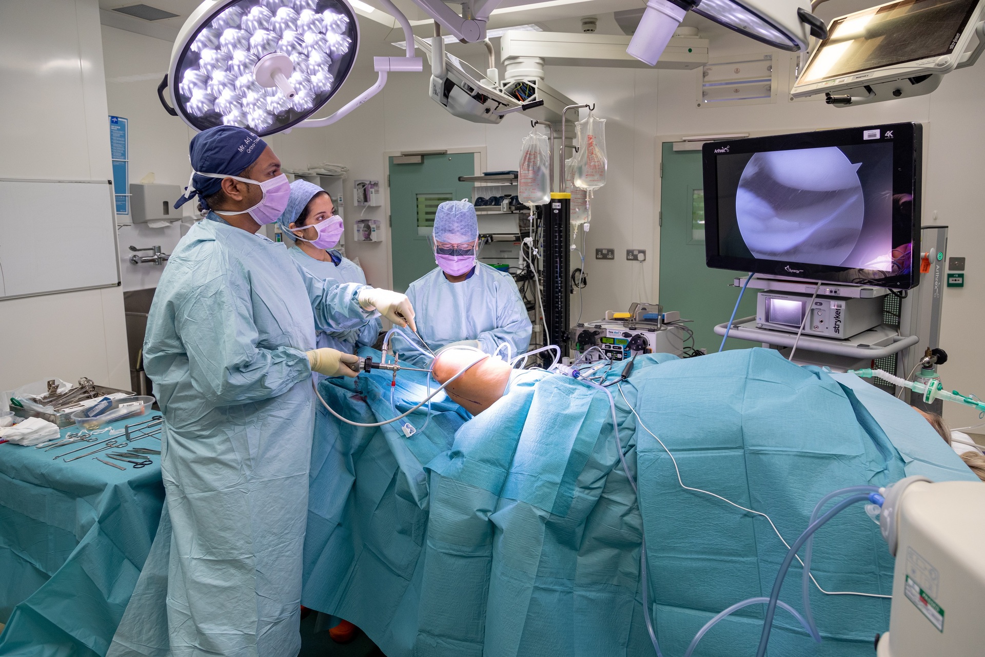
The surgeon will then carefully make two small 1cm incisions, called ‘portals’, on your knee, in the locations shown on the image below. The knee joint will then be filled with a sterile saline solution to clear gently create some space in the knee between the tibia and femur bone for the instruments and constantly flush the knee off any debris or blood.
This will also make the image produced by the arthroscope clearer. The precise placement of these portals is important, as the joint space is quite narrow, so accidental damage to the meniscus or cartilage of the joint can potentially occur and ideally the second portal should be made under direct vision via the arthroscope which is placed in the knee through the first portal.
Once the arthroscope has been inserted through the portals into the knee joint space, structures in the knee are thoroughly and systematically examined to inspect for further damage. These structures include the anterior and posterior cruciate ligament, articular cartilage, Hoffa’s fat pad, synovial plicae and both the medial and lateral menisci.
The meniscus tear is then identified and probed to assess the size, location, stability, tissue quality and pattern of the tear. At this stage, the decision is made whether to repair or remove the damaged meniscus tissue. If the meniscus is red and vascular as opposed to white and avascular it should be amenable to repair.
The two sides of the torn meniscus tissue are then prepared for healing by using a small motorised shaver or even a long thin needle to pierce the fragments and promote bleeding.
If the blood supply to the meniscus is damaged, several techniques are available to improve it. These have variable scientific evidence to support them but include placing a blood clot between the tear itself, creating small blood vessels around the edges of the meniscus or causing a controlled bleed in the joint lining.
To complete the procedure, the meniscus is sewn into place using various types of suture devices.
There are a number of different described techniques to repair a meniscus and they are named according to the direction of travel of the specialised arthroscopic needles.
The most common currently is the all inside technique using hybrid systems of sutures attached to small tags which sit behind the meniscus. The all inside technique is best for tears of the posterior part of the meniscus at the back of the knee. The outside in technique is best for anterior tears at the front of the meniscus, whilst the inside out technique is less common now as it is technically demanding and requires more incisions.
Once the repair is performed and the fixation deemed solid, the surgical tools are removed from the knee and the small portals are closed with suture or paper stitches or skin glue according to your surgeon’s preference.
Waterproof dressings are then placed over the knee and a compression bandage for 12-24 hours to help reduce any swelling.
The patient must keep the knee dry for 2 weeks till your follow up appointment with your surgeon where the skin incisions will be checked to ensure they have healed, and any stitches removed.
If a meniscal repair is performed, you will need to wear a special hinged knee brace when walking for 6 weeks. This prevents excessive knee bend while the meniscus is trying to heal and limits the risk of failure of the repair.
If the torn meniscus is removed, you will not need a knee brace.
Post-operative surgery care after your procedure
Rehabilitation post-operatively aims to reduce swelling, improve range of movement and strengthen the quadriceps (thigh) muscles. You will be seen by a physiotherapist before and ideally after your discharge from the hospital. The physiotherapist will show you exercises designed to strengthen your knee and prevent it from becoming stiff.
These exercises should be performed immediately to improve range of motion and strength in the leg muscles. You may begin practising standing on one leg once pain and swelling has subsided. To help ease into improving your balance, you may start by using the wall as a support, and then gradually transition to standing on one leg unassisted.
You will be provided with crutches and taught how to use them by the physiotherapist, with full weightbearing allowed as you can tolerate.
You should return your crutches to the physiotherapy department or to your surgeon in a follow up appointment once you no longer need them.
If you experience persistent pain, swelling, oozing from the wound, fever or calf pain, it is essential that you contact your GP immediately or attend the Urgent Care Centre or Emergency Department linked to the hospital your surgeon works in or where the operation was performed for evaluation.
ACL Reconstruction
Hear Mr Imbuldeniya explain what an ACL injury is and how to recover if you need surgery.
Just 11 days after surgery, Millie's wounds have healed, she's hardly got any swelling, and is beginning the road to recovery with our Physiotherapists.
ACL injuries: what you need to know
What is the anterior cruciate ligament?
The anterior cruciate ligament (ACL) is one of the four main ligaments in the knee which work together to provide knee stability (the others are the posterior cruciate ligament [PCL], medial collateral ligament [MCL] and lateral collateral ligamentt [LCL]). The ACL is located in the middle of the knee and runs diagonally from the back of the femur (thigh bone) to the front of the tibia (shin bone). It prevents the tibia from moving excessively forward in relation to the femur and stops inward rotation of the knee.
What causes anterior cruciate ligament injury?
Injuries to the ACL include sprains and complete or partial tears, although complete tears are the most common.
ACL injuries usually occur as a result of twisting or pivoting injuries to the knee, such as stopping and changing direction quickly whilst running or jumping. Because of this, ACL injuries are common in sports such as football, basketball, tennis, volleyball and skiing which involve turning and twisting movements. The ACL can also be torn following direct impact to the knee during contact sports such as rugby, but this is less common.
ACL injuries are generally more common in women than in men due to differences in knee joint anatomy, muscle bulk and because the female hormone oestrogen affects joint laxity.
What are the symptoms of anterior cruciate ligament injury?
Most people hear and/or feel a ‘popping’ noise in their knee when they injure their ACL. There is also usually immediate pain especially on weight-bearing, and you may experience swelling within the first 6 hours, a reduced range of movement in the knee and feeling that the joint is unstable (‘giving way’ whilst walking or during physical activity).
Can you still walk if you have a torn anterior cruciate ligament?
Yes. Some people can live and function normally with a torn ACL, however most people find it causes their knee to feel painful or unstable and they may have difficulty walking, running or ascending/descending stairs on their injured leg.
How do you treat an anterior cruciate ligament injury?
Initial first aid treatment for a torn ACL includes resting the knee, applying ice and compression to it to and taking anti-inflammatory and pain-relieving medications to help reduce pain and swelling. Using crutches whilst walking can also help to keep the weight off your knee.
It is recommended that you see an orthopaedic specialist as soon as possible to confirm the diagnosis and give you appropriate treatment options. You are likely to require tests such as an MRI scan to help with this.
Not all torn ACLs require surgery; this will depend on whether you have symptoms of instability (‘giving way’ of the knee) or if you like to participate in sports.
Surgery is usually required if you hope to get back to physical activity, and can either be performed immediately after injury or a while down the line to allow the swelling and inflammation in the knee to settle.
Whether you are planning to have surgery or not, physiotherapy is important in ACL rehabilitation. Mobility and strengthening exercises help to improve muscle bulk around the knee in order to improve its stability, and to get your range of movement back to normal.
If you are going to have surgery on your torn ACL, it is important to have physiotherapy beforehand to optimise your knee muscles and movements in order to get the best results from your operation: this has been shown to improve overall outcomes.
The definitive management for a torn ACL with instability is reconstructive surgery, where your surgeon removes the damaged ACL and replaces it with tissue to help a new ligament grow in its place. This tissue usually comes from either your hamstring tendons or patellar tendon, although sometimes artificial or donor ligaments can be used instead. The tissue is held in place at either end using bony plugs, screws or other fixation devices.
Can the anterior cruciate ligament repair itself?
No. Unlike torn muscles, a torn ACL cannot heal by itself even though your knee may feel better after a couple of weeks of rest.
Returning to physical activity without having it checked risks re-injury and damage to other structures in the knee, such as the meniscus or other ligaments.
After surgery you will be able to walk without crutches by around 6 weeks, and with regular physiotherapy patients can often get back to sports again within a year after their operation.
Knee Arthroscopy
What are the advantages of arthroscopy?
Arthroscopy offers some advantages over traditional open surgery:
High-definition images of your joint
Small surgical instruments can be accurately inserted
Less pain after the operation
Faster recovery time
Lower risk of infection
Most patients can go home the same day
You may be able to return to daily activity more quickly
Who would benefit from arthroscopy?
You might need an arthroscopy if you have persistent joint pain, catching, swelling or stiffness. An arthroscopy may be used to examine the degree of joint damage caused by injury, such as a sports injury or from conditions that can cause joint damage, such as osteoarthritis.
The operation can
be used to treat joint problems and conditions:
Damaged cartilage
Removing fragments of loose bone or cartilage.
Draining away fluid
Treating arthritis or carpal tunnel syndrome
Treatment facts
An arthroscopy usually takes around an hour to perform depending on what needs to be done.
You will usually have a general anaesthetic, but it is possible for an arthroscopy to be done under local anaesthetic while you’re awake. This means you’ll be able to go home straight afterwards. If you’re going under a general anaesthetic, you’ll need to stop drinking and eating at least six hours prior to the procedure.
During the operation, your orthopaedic surgeon will make small incisions around the affected joint. Sterile fluid will then be pumped into the joint so that the camera can collect a clearer picture.
Then the arthroscope will be inserted into the incisions and the injury will be examined by looking at the images that are sent to the monitor attached to the arthroscope.
If your surgeon is repairing a joint, he or she will insert small instruments through a small separate incision.
Recovery
Recovery time depends on the injured joint and the specific operation. Patients usually return to light, physical activities within a few weeks, but more strenuous activities such as lifting and sport may be delayed for several months.
Our dedicated physiotherapy team will help you get back on your feet using The ICE Ortho Clinic’s exclusive patient rapid recovery pathway,
Risks
An arthroscopy is widely considered to be a safe procedure, though it does carry some risks. It’s normal to experience temporary problems such as swelling, bruising, stiffness and discomfort after an arthroscopy. Your surgeon will discuss with you the possible risks.
Hyaluronic acid injections
Sodium hyaluronate (also known as hyaluronic acid or hyaluronan) is a natural substance. It is present in the body wherever moisture is stored or lubrication between layers of tissue is required to eliminate friction.
Examples are inside the eye ball itself, the tear film, the joint cartilage, the synovial fluid in the joints, all the mucuous membranes of the body, but also the basic substance of the skin which consists of up to 55% sodium hyaluronate.
Sodium hyaluronate takes on a variety of functions:
Storage: it absorbs and stores moisture in enormous quantities
Lubrication
Transport medium for nutrients
Filter for inflammatory molecules
It may help to visualise sodium hyaluronate as a dish of spaghetti (this is what the molecules look look like under an electronic microscope): a dense mesh that remains elastic and flexible in spite of its density and interwoven structure.
Sodium hyaluronate is an essential component of the synovial fluid. It enables the fluid to act as a lubricant, a shock absorber and a filter controlling the movement of cells and large molecules within the joint.
Sodium hyaluronate is injected into the space in the joint that contains synovial fluid and works by restoring the normal balance between the breakdown and production of sodium hyaluronate. This procedure is known as 'viscosupplementation.'
One such brand used in our clinic is OSTENIL® PLUS which can decrease pain and stiffness and improve the other symptoms of osteoarthritis.
The sodium hyaluronate in OSTENIL®PLUS is very pure and is manufactured using a process called fermentation. It contains no animal proteins, which means that it is very unlikely to cause an allergic reaction. OSTENIL® PLUS has been given to thousands of patients and has not been found to cause any serious side effects. The exact make-up of the sodium hyaluronate in OSTENIL®PLUS has been carefully chosen so that it is as effective as possible in treating osteoarthritis.
Only one injection per joint is required, with pain relief often lasting for many months.
Arthrosamid®
Arthrosamid® is a novel treatment offered by Mr Imbuldeniya and the West London Knee and Hip Clinic for knee osteoarthritis. It offers an effective, safe, long lasting, non surgical, injectable alternative to existing therapies. This non-biodegradable hydrogel is injected under precise ultrasound guidance into the knee joint to alleviate pain and cushion the joint.
It's a minimally invasive outpatient procedure, ensuring you don't need to stay in the hospital. Arthrosamid® is safe and biocompatible, integrating seamlessly with knee tissues, backed by two decades of research and approx. 1 million units used for various indications.
In clinical trials, patients experienced reduced pain levels by Week 4 post-injection, and this relief was sustained for up to 156 weeks (3 years) in some cases. In general, most (80%) of patients have significant, though not total pain relief for 1-2 years. The trials will continue for five years.
Knee osteoarthritis is a chronic condition causing joint pain and stiffness, typically treated with exercise, weight loss, pain relief medication, viscosupplement injections, PRP injections, anti-inflammatory corticosteroid injections, supportive knee braces, and physiotherapy. Total knee replacement is the last resort. However, Arthrosamid® offers a promising change.
Arthrosamid® is a 2.5% cross-linked polyacrylamide hydrogel that, when injected, cushions the knee joint, providing safe and sustained relief with a single, one off injection. It restores viscosity in the synovial fluid, improving joint lubrication and cushioning, and integrates into the synovium of the inner joint capsule.
This treatment reduces pain, stiffness, and improves mobility, enhancing your quality of life.
Platelet rich plasma injections
PRP therapy (Platelet Rich Plasma therapy), also known as Autologous Conditioned Plasma (ACP) therapy, takes advantage of the blood’s natural healing properties to repair damaged cartilage, tendons, ligaments, muscles, or even bone. The main aim of this treatment is to reduce pain, improve joint function and potentially slow, halt or even repair damage to cartilage. It is well used in Medicine and helps patients regain activity by effectively reducing pain and improving mobility. This simple outpatient treatment may help you recover from a painful sports injury or chronic pain caused by osteoarthritis and tendonopathy.
The procedure
The procedure is performed in the out-patient clinic.
A blood sample is drawn from a vein in the arm as per any regular blood test you might have had before.
Your blood sample is placed in a special machine which spins the blood in a centrifuge to seperate, extract and concentrate the desired substances (ACP Autologous Conditioned Plasma)
These substances (ACP) are injected back into your painful knee or hip or the affected tendons nearby.
The treatment is generally administered at three weekly intervals (3 treatments in total). Since the treatment is prepared from the patient’s own blood, the treatment carries a very low risk.
How it works
The healing of injured or inflamed tissue involves a complex and precisely regulated series of natural processes within the body. Thrombocytes (platelets) play an important role in this process. At the site of injury they release growth factors that initiate the restoration of injured tissue and inhibit painful inflammatory processes. PRP therapy is based on our understanding of these complex processes. With its high concentration of growth factors PRP injections are a natural way of supporting the body’s self-healing processes.